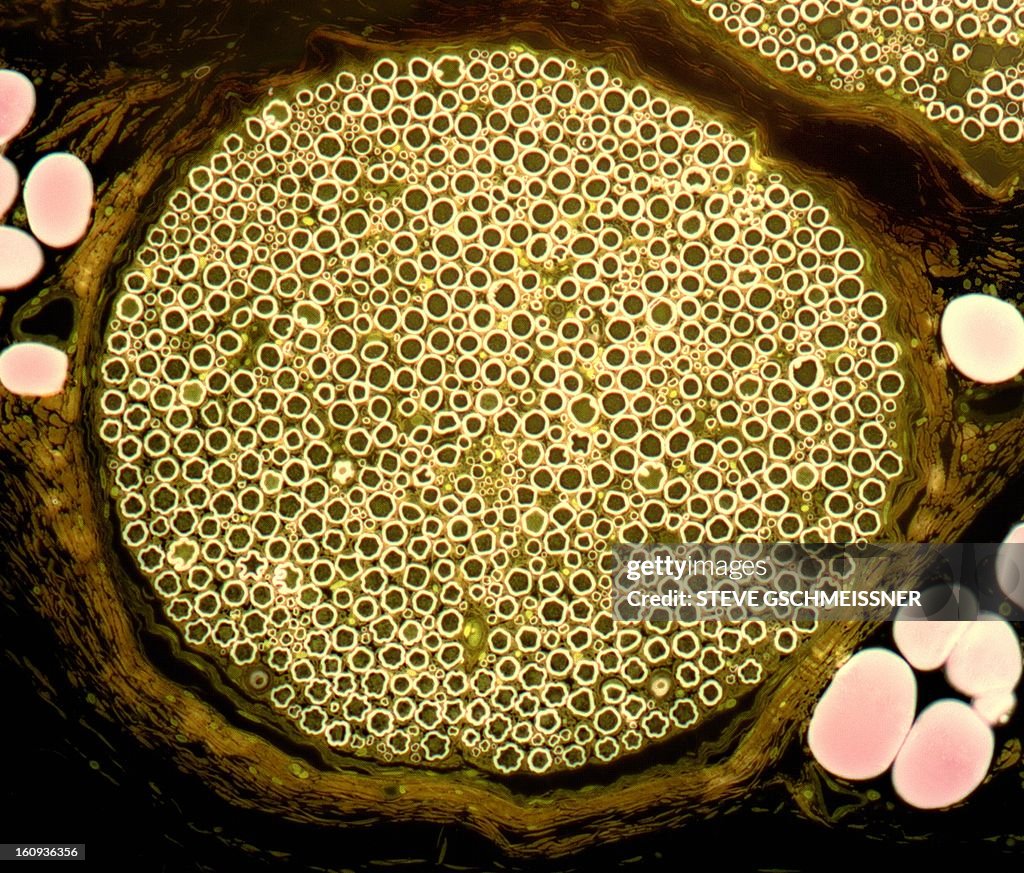
Nerve bundle, light micrograph - stock illustration
Nerve bundle. Light micrograph of a section through a nerve bundle from the sciatic nerve. Myelin sheaths (bright circles) can be seen surrounding the axons (dark dots). Perineurium (connective tissue, brown) surrounds the nerve bundle. Adipose (fat) cells (pink) surround the nerve bundle. Magnification: x200 when printed at 10 centimetres wide.

Get this image in a variety of framing options at Photos.com.
PURCHASE A LICENSE
All Royalty-Free licenses include global use rights, comprehensive protection, simple pricing with volume discounts available
kr 2,500.00
NOK
Getty ImagesNerve Bundle Light Micrograph High-Res Vector Graphic Download premium, authentic Nerve bundle, light micrograph stock illustrations from Getty Images. Explore similar high-resolution stock illustrations in our expansive visual catalogue.Product #:160936356
 Download premium, authentic Nerve bundle, light micrograph stock illustrations from Getty Images. Explore similar high-resolution stock illustrations in our expansive visual catalogue.Product #:160936356
kr2,500kr300
Getty Images
In stockDETAILS
Credit:
Creative #:
160936356
License type:
Collection:
Science Photo Library
Max file size:
4572 x 3901 px (15.24 x 13.00 in) - 300 dpi - 9 MB
Upload date:
Release info:
No release required
Categories:
- Anatomy,
- Art Product,
- Biological Cell,
- Biology,
- Biomedical Illustration,
- Circle,
- Color Image,
- Digitally Generated Image,
- Healthcare And Medicine,
- Horizontal,
- Human Internal Organ,
- Human Nervous System,
- Illustration,
- Large Group Of Objects,
- Magnification,
- Microbiology,
- Myelin Sheath,
- Nerve Bundle,
- No People,
- Sciatic Nerve,
- Science,
- Scientific Micrograph,