SPINAL CORD. CROSS SECTION, 2.5X. Shows: gray matter, white matter, dorsal root ganglia, dorsal and ventral roots, central canal, anterior horn cells (motor neuron cell bodies) & meninges - stock photo

Get this image in a variety of framing options at Photos.com.
PURCHASE A LICENSE
All Royalty-Free licenses include global use rights, comprehensive protection, simple pricing with volume discounts available
kr 2,500.00
NOK
Getty ImagesSpinal Cord Cross Section 25x Shows Gray Matter White Matter Dorsal Root Ganglia Dorsal And Ventral Roots Central Canal Anterior Horn Cells Meninges High-Res Stock Photo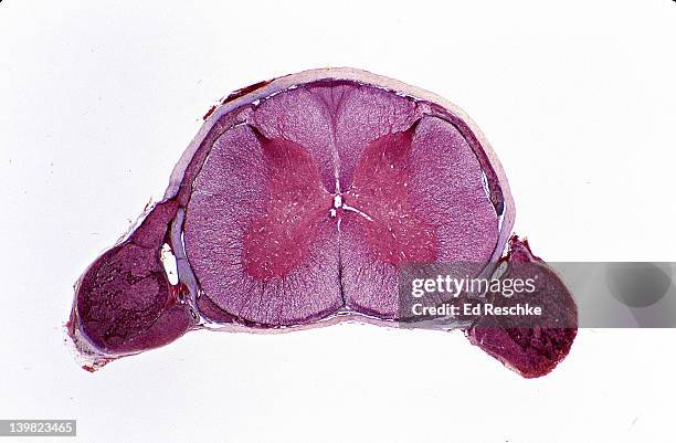 Download premium, authentic SPINAL CORD. CROSS SECTION, 2.5X. Shows: gray matter, white matter, dorsal root ganglia, dorsal and ventral roots, central canal, anterior horn cells (motor neuron cell bodies) & meninges stock photos from Getty Images. Explore similar high-resolution stock photos in our expansive visual catalogue.Product #:139823465
Download premium, authentic SPINAL CORD. CROSS SECTION, 2.5X. Shows: gray matter, white matter, dorsal root ganglia, dorsal and ventral roots, central canal, anterior horn cells (motor neuron cell bodies) & meninges stock photos from Getty Images. Explore similar high-resolution stock photos in our expansive visual catalogue.Product #:139823465kr2,500kr300
Getty Images
In stockDETAILS
Credit:
Creative #:
139823465
License type:
Collection:
Stone
Max file size:
4443 x 2911 px (14.81 x 9.70 in) - 300 dpi - 7 MB
Upload date:
Release info:
No release required
Categories:
- Cross Section,
- Spine - Body Part,
- Scientific Micrograph,
- Front View,
- Biological Cell,
- Canal,
- Car Horn,
- Color Image,
- Distorted,
- Engine,
- Gray Color,
- Growth,
- Horizontal,
- Human Body Part,
- Human Bone,
- Human Brain,
- Human Internal Organ,
- Human Spine,
- Immunofluorescent Photomicrograph,
- Magnification,
- Material,
- Meninges,
- Microscope,
- Nerve Cell,
- Photography,
- Research,
- Science,
- Scrutiny,
- Showing,