Muscle fibre, SEM - stock video
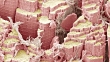
Muscle fibre. Coloured scanning electron micrograph (SEM) of a freeze-fractured skeletal (or striated) muscle fibre. The fracturing of the fibre has revealed that it consists of a bundle of smaller fibres called myofibrils. The myofibrils are crossed by transverse tubules (horizontal lines), that mark the division of the myofibrils into contractile units (sarcomeres). Magnification: x8000 when printed at 10 centimetres wide.


PURCHASE A LICENSE
Get personalized pricing by telling us when, where, and how you want to use this asset.
DETAILS
Credit:
Creative #:
618321807
License type:
Rights-ready
Collection:
Image Bank Film
Max file size:
1920 x 1080 px - 550 MB
Clip length:
00:00:12:00
Upload date:
Release info:
No release required
Mastered to:
QuickTime 8-bit Photo-JPEG HD 1920x1080 25p
Categories:
- Muscle,
- Anatomy,
- 10 Seconds or Greater,
- Art Product,
- Biology,
- Biomedical Illustration,
- Color Image,
- Colors,
- Coral Colored,
- Digitally Generated Image,
- Film - Moving Image,
- HD Format,
- Horizontal,
- Human Body Part,
- Human Skeleton,
- Ideas,
- Magnification,
- Microbiology,
- Muscle Fiber,
- Myofibril,
- No People,
- Real Time Video,
- SEM,
- Sarcomere,
- Scanning Electron Microscope,
- Science,
- Striped,
- Tubules,
- Zoom Out,